Ultrasound is a procedure of imaging that is non-invasive to examine organs, tissues, or unborn children. It is done by high-frequency sound waves in place of radiation and hence can be applied to almost all diagnostic and monitoring applications, since it is safe. Whether ultrasound is used to scan the belly or detect problems with vascular blood flow, the technology plays a vital role in diagnosis nowadays. This guide teaches how ultrasounds are performed, the various types, the reasons, and what to expect before, during, and after the process.
What Is an Ultrasound?
Having a medical imaging form, sonography (USG: u/s) or ultrasound is based on high-frequency sound waves, producing images of the inside of the body. Also called sonograms, these images are used by doctors to study such organs as the liver, kidneys, bladder, or heart or to track the progression of pregnancy. Contrary to X-rays, ultrasounds do not involve ionizing radiation; hence, they can be repeated.
The ultrasound equipment consists of a probe (transducer) that transmits sound waves into the body. The tissues reflect the waves off them, producing echoes that are converted into picture-like images on a monitor. The resolution of such images is dependent upon the kind of ultrasound as well as the region under consideration.
How Does an Ultrasound Work?
The ultrasounds employ the law of reflection of sound waves. The transducer carries high-frequency sound waves that pierce the body and bounce back upon hitting tissues or organs. The machine measures these echoing returns and computes them into a two-dimensional or a three-dimensional image.
The intensity and velocity of echoes rely on the density of the tissues. As an example, bones can reflect a higher number of sound waves, whereas fluids can transmit sound. The comparison helps to create realistic pictures. The kind of sound wave applied—which is the frequency and the depth—changes according to the part of the scanned body.
Types of Ultrasounds
2D, 3D, and 4D Ultrasound
Ordinary ultrasound makes two-dimensional black and white images called 2D ultrasound. It is the most widespread one, and it is employed in abdominal scans and fetal scans.
3D ultrasound gives depth because it integrates several 2D images to create a 3D image. It aids in a better identification of structural deviations, particularly in pregnancy.
The 4D ultrasound incorporates movement into the 3D images. It is mostly applied to see real-time fetal movement and expressions across the face. The 4D scans can be useful to diagnose some prenatal disorders, though they are frequently requested by pregnant parents.
Doppler Ultrasound
This form of ultrasound involves assessment of blood circulation in veins and arteries. It aids in the identification of blockage, clots, and unusual flow and is therefore crucial in the diagnosis of medical conditions in the cardiovascular system. A Doppler ultrasound is a measurement of both the direction and speed of blood using the frequency change of the dominant sound waves.
Abdominal Ultrasound
Applied in the study of the liver, gallbladder, kidneys, pancreas, and other organs of the abdomen. It allows diagnosing diseases such as gallstones, kidney stones, and liver disease.
Pelvic and Transvaginal Ultrasound
They are applied to evaluate the uterus, ovaries, and bladder. Transvaginal ultrasound gives a closer examination of the reproductive organs of women, and it is commonly applied in such cases in early pregnancy or the assessment of unexpected pelvic pain.
Transrectal and Prostate Ultrasound
They are geared towards looking at the prostate gland. A probe (small probe) is inserted into the rectum to receive detailed images, which are normally applied in procedures meant to diagnose prostate conditions.
Neck and Thyroid Ultrasound
Neck ultrasound is used to analyze the lumps, nodules, or swellings in the thyroid glands or lymph nodes. It offers a radiation-free approach to evaluate the thyroid activity and anatomy.
Sonogram vs. Ultrasound vs. Sonography
These terms are often interchanged, although they slightly differ. Ultrasound is the imaging, sonography is a practice or technique, and lastly, is sonogram is the result or picture produced. Learning about this difference will assist with reading medical reports and directions.
Who Performs an Ultrasound?
Ultrasounds are usually administered by special medical equipment known as a sonographer or an ultrasound technologist. These experts will be trained on how to use the ultrasound machine, obtain superior images, and ensure the safety of the patient during the process. In other instances, interpretation of the scan may also be conducted by the doctors: a radiologist, obstetrician, or cardiologist.
Sonographers have to be formally educated and clinically trained. They are knowledgeable in anatomy, the handling of equipment, and image-interpretation basics. The salaries of ultrasound technicians are dependent on their qualifications, location, and specialization.
Why an Ultrasound Is Done
Pregnancy Monitoring
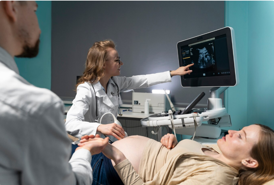
The best-known ultrasound usage is when it is related to pregnancy. It assists in confirming pregnancy, monitoring fetal growth, diagnosing birth defects, estimating gestational age, and monitoring the baby’s motions through 3D or 4D images of the ultrasound. Level 2 scans are a close examination of the fetus regarding anatomy.
Disease Diagnosis
The ultrasound is applied in the diagnosis of different complications in the abdomen, pelvis, neck, heart, and other body areas. It is able to detect cysts, tumors, fluid accumulation, flow of blood, and swelling. It also comes in handy when evaluating liver, kidney, gallbladder, and thyroid issues.
Guiding Medical Procedures
Instant ultrasound imagery helps in positioning the needle in biopsies, fluid removal, and even in some operations. It is used to aid the safety and accuracy of procedures such as injections or inserting a catheter.
Preparing for an Ultrasound
Depending on the type of scan, the preparation is made differently. With abdominal scans, the patient may need to fast 68 hours before to empty the body of gas that will block the visibility of images. In the case of a pelvic or bladder scan, you might be expected to drink water and keep a full bladder to ensure a better visualization.
Do not wear tight clothes, jewelry, etc., around the region of scanning. You might be requested to wear a gown. The skin around the exam area should also not be covered with lotions or powders.
What Happens During the Procedure
In ultrasound, a healthcare provider will require the patient to lie on a table with some water-based gel being spread on the skin covering the part that is being investigated. This gel makes the sound waves conduct effectively.
The sonographer will use a transducer on the skin, and she will press it to get good pictures. The scan game is usually 15 to 45 minutes long, with the amount based on complexity. However, in other situations, such as transvaginal or transrectal ultrasounds, the probe is inserted to have a better picture, which may be a bit uncomfortable but is not normally painful.
Are Ultrasounds Painful or Risky?
The process of ultrasound, as a rule, is not painful. You can only experience some slight strain or pain when undergoing internal scans. It is also among the safest imaging tools out there, particularly without radiation exposure. It is 100 percent safe among pregnant women and is being used all over the world to assess fetuses.
Understanding Ultrasound Results
A radiologist or a specialist normally reviews the results of an ultrasound. They make meaning of the images and send a comprehensive report to your doctor, who has referred you. This report could contain the measurements that had been done, observations collected, and any probable abnormalities that had been identified.
Your doctor can prescribe additional tests, scans, or treatment, depending on the results. In case any indistinct image is seen in the pictures, further imaging such as CT or MRI may be prescribed.
Common Questions to Ask About Your Ultrasound
- What are you looking for in this scan?
- How accurate are the results?
- When and how will I receive my report?
- Will I need another scan later?
- Is any special care needed after the procedure?
Conclusion
Ultrasound In Las Vegas is another very effective and multi-purpose diagnostic instrument with detailed images of internal body organs, blood vessels, and fetal development that does not involve the risk of radiation. Whether going through a regular pregnancy scan, checking abdominal discomfort, or following up on a health condition, the knowledge of how it happens may help reduce the anxiety and enhance effective communication with the provider.
You should pay attention to the preparation instructions in case you plan to undergo an ultrasound. When you see your results, remember not to be afraid of asking questions; effective communication means improved health conditions.
To schedule your appointment with the highest standard of care and visit Sahara West Urgent Care, visit our website.
Faqs
Is ultrasound better than an X-ray?
They serve different purposes. Ultrasound is better for soft tissues and is safer for repeated use, especially during pregnancy.
Can ultrasound detect all diseases?
No. While it can detect many conditions, some issues may require further imaging, like MRI or CT scans.
Is there any recovery time after an ultrasound?
No recovery time is needed. You can resume normal activities immediately after the scan.
How many ultrasounds are done during pregnancy?
It varies. Typically, at least two are done—one early on and another around 20 weeks. More may be required based on medical needs.
Are 3D and 4D ultrasounds necessary?
They are not medically necessary in most cases, but may be useful if your doctor needs detailed images of the fetus.
